Gum Biopsy in Brooklyn
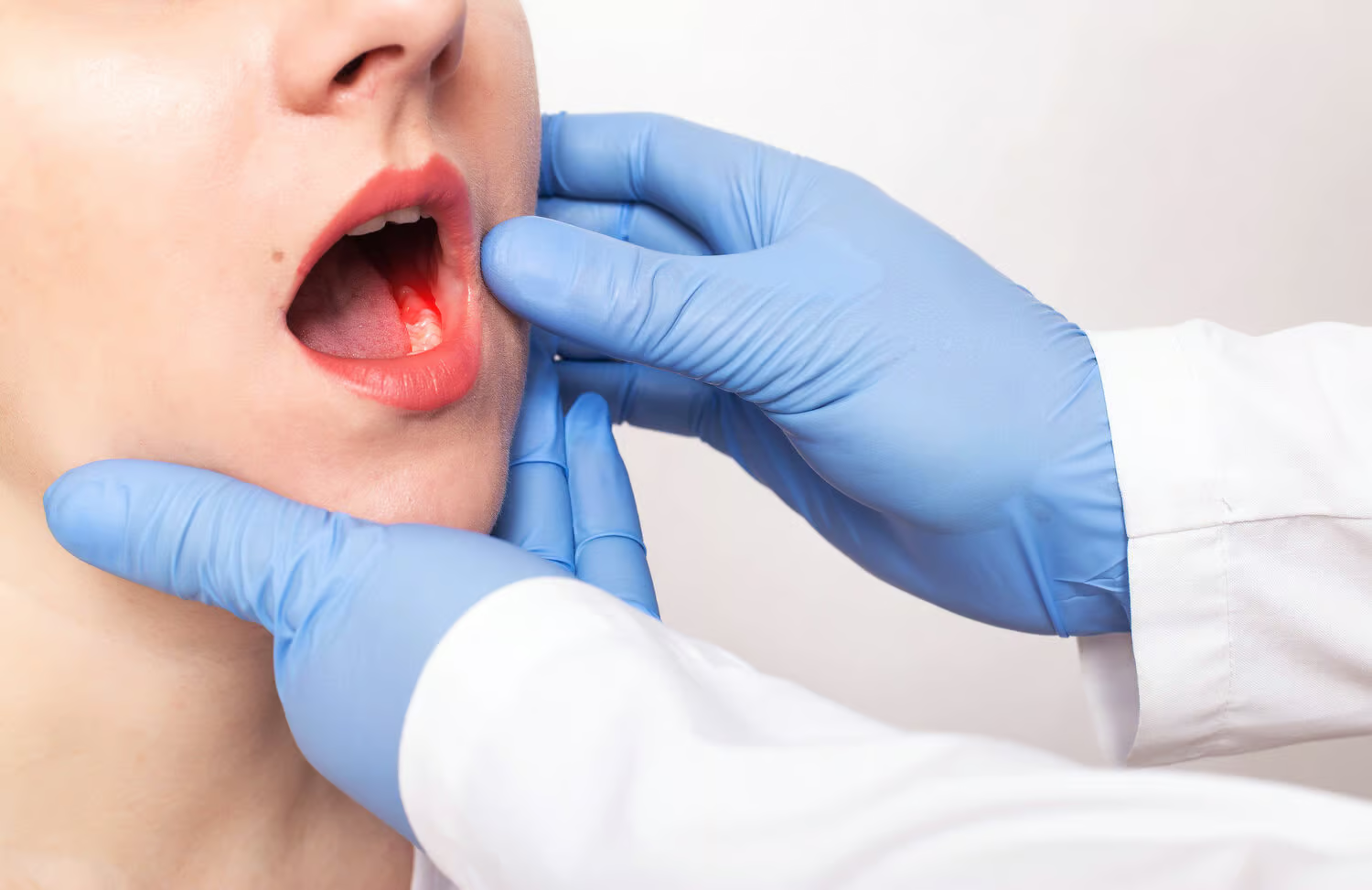
When your dentist does an examination, he looks for abnormal gum tissue as well as cavities in your teeth. If he finds unusual or abnormal gum tissue, he may perform a gum biopsy. During a gum biopsy, your dentist removes a tiny amount of tissue from your gums. Then he sends it to a lab for testing to determine if you have oral cancer or non-cancerous growths in your mouth.
Types of Gum Biopsies
Incisional Biopsies
Incisional biopsies occur when a dentist removes a tiny amount of tissue and examines it under a microscope. A pathologist also looks at the tissue to determine a diagnosis regarding the tissue sample.
Excisional Biopsies
During an excisional biopsy, your dentist removes an entire lesion. This lesion is removed along with some surrounding healthy tissue, then examined for cancer cells.
Percutaneous Biopsies
When performing a percutaneous biopsy, the doctor inserts a needle into the questionable area to remove cells. The cells are then tested, and a diagnosis made. A fine needle biopsy or a core needle biopsy may be done, depending on the doctor’s preference.
Brush Biopsy
During a brush biopsy, a brush is rubbed against the area of concern in the mouth. The skin cells collected from the brush are tested for signs of cancer. If cancer is detected, further testing occurs to learn more about the cancer. Brush biopsies remain the most frequently used form of initial diagnosis for oral cancers.
Frequently Asked Questions About Oral Biopsies
Is an oral biopsy painful?
No. We use local anesthesia and gentle techniques. Most patients feel only mild soreness afterward.
How long does the biopsy take?
The procedure typically takes 15–30 minutes.
What should I expect after the biopsy?
Mild discomfort, swelling, or sensitivity for a day or two. Most people resume normal eating and activity quickly.
Does a biopsy mean I have cancer?
Not necessarily. Most biopsies are precautionary. Early diagnosis, when necessary, allows for better outcomes.
Is a biopsy covered by insurance?
Yes, in most cases. Our team will help explain your insurance coverage and financing options.
Lorem ipsum dolor sit amet, consectetur adipiscing elit, sed do eiusmod tempor incididunt ut labore et dolore magna aliqua. Ut enim ad minim veniam, quis nostrud exercitation ullamco laboris nisi ut aliquip ex ea commodo consequat. Duis aute irure dolor in reprehenderit in voluptate velit esse cillum dolore eu fugiat nulla pariatur.
Schedule an Oral Biopsy Evaluation in Brooklyn
If you’ve noticed a sore or spot in your mouth that doesn’t heal, don’t wait to get it checked. At Shawn Cohen D.D.S. P.L.L.C. on Avenue U in Brooklyn, we offer gentle, thorough evaluations and expert biopsy services to help you stay proactive with your oral health.
Call (718) 372-3151 or book your consultation online today.